

Skull Anatomy Quick Guide: Bones, Muscles & Labeled Diagram
The skull is much more than the bone that gives the head its shape. It protects the brain, supports the face, anchors the muscles used for chewing and facial expression, and houses important structures involved in vision, hearing, breathing, and speech. Because so many essential functions depend on it, even minor injuries or abnormalities can affect daily life.
Many people only think about skull anatomy after a facial injury, persistent jaw pain, or a recommendation for imaging following an accident. Medical terminology and anatomy diagrams can feel overwhelming, making it difficult to understand which bones are involved, how they fit together, or why certain injuries require specialized treatment.
Learning the basic anatomy of the skull can make conversations with your healthcare provider easier and help you better understand diagnoses, imaging results, and treatment recommendations. Whether you're curious about the bones of the face, the muscles that move the jaw, or how the skull protects the brain, having a clear overview provides valuable context.
This guide explains the major parts of the skull, identifies its primary bones and muscles, explores what's inside the skull, reviews common injuries and diagnostic tests, and answers frequently asked questions about skull anatomy and its clinical importance.
What Is the Skull?
The skull is the bony framework of the head that protects the brain and supports the facial structures. It is composed of 22 bones that are connected by strong, immovable joints called sutures, except the lower jaw (mandible), which forms a movable joint with the skull.
The skull is typically divided into two major regions:
- The cranium, which encloses and protects the brain.
- The facial skeleton, which forms the framework of the eyes, nose, cheeks, and jaws.
Together, these bones create a rigid yet lightweight structure that safeguards vital organs while providing attachment points for muscles involved in chewing, speaking, swallowing, and facial expression.
Although the skull appears to be a single bone, it is actually a complex arrangement of individual bones that fuse during growth and development.
What Is the Function of the Skull?
According to the National Library of Medicine (NIH), “its purpose is to protect the brain and allow attachments for the facial muscles.”
Given that, we can go deeper and say that the skull performs several essential functions that support both protection and normal daily activities.
Its primary functions include:
- Protecting the brain from injury.
- Supporting the facial structures, including the eyes, nose, and mouth.
- Housing sensory organs responsible for vision, hearing, smell, taste, and balance.
- Providing attachment points for muscles that control chewing, swallowing, facial expressions, and head movement.
- Forming passages for nerves and blood vessels that travel between the brain and the rest of the body.
- Helping maintain the shape of the head and face, contributing to both appearance and function.
Because many muscles, nerves, and joints interact with the skull, injuries affecting its bones can influence speaking, eating, breathing, vision, and other important functions.
What Are the Main Parts of the Skull?
The skull is made up of numerous bones, muscles, joints, and soft tissues that work together to protect the brain and support essential functions such as chewing, speaking, breathing, and facial expression. Understanding these structures makes it easier to interpret medical imaging, understand injuries, and discuss treatment options with healthcare providers.
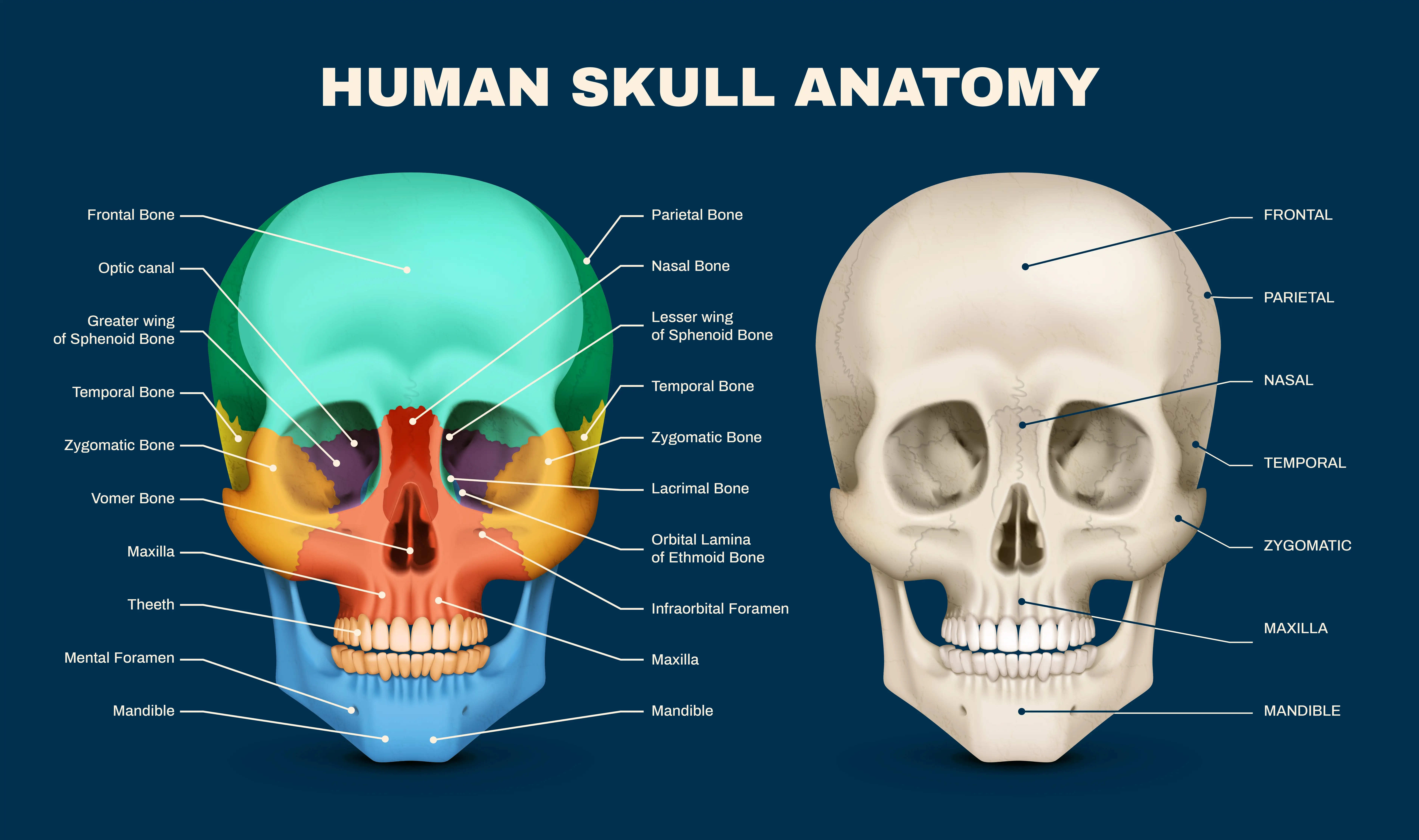
Skull Bones
The adult skull consists of 22 bones, divided into two groups:
Cranial bones (8):
- Frontal bone
- Parietal bones (right and left)
- Temporal bones (right and left)
- Occipital bone
- Sphenoid bone
- Ethmoid bone
These bones form the cranium, the protective case surrounding the brain.
Facial bones (14):
- Maxillae (right and left)
- Zygomatic bones (right and left)
- Nasal bones (right and left)
- Lacrimal bones (right and left)
- Palatine bones (right and left)
- Inferior nasal conchae (right and left)
- Vomer
- Mandible
Together, these bones provide the framework for the face, support the teeth, and create the openings for the eyes, nose, and mouth.
Skull Muscles
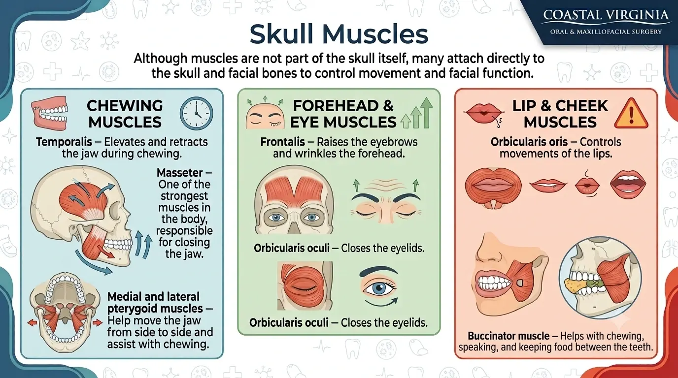
Although muscles are not part of the skull itself, many attach directly to the skull and facial bones to control movement and facial function.
Some of the most important muscles include:
- Temporalis – Elevates and retracts the jaw during chewing.
- Masseter – One of the strongest muscles in the body, responsible for closing the jaw.
- Medial and lateral pterygoid muscles – Help move the jaw from side to side and assist with chewing.
- Frontalis – Raises the eyebrows and wrinkles the forehead.
- Orbicularis oculi – Closes the eyelids.
- Orbicularis oris – Controls movements of the lips.
- Buccinator – Helps with chewing, speaking, and keeping food between the teeth.
These muscles work together with the facial bones and temporomandibular joints (TMJs) to perform everyday functions.
Skull Labeled Diagram
A labeled skull diagram helps visualize how the different bones fit together and where important anatomical landmarks are located. Most diagrams identify major structures such as the frontal bone, parietal bones, temporal bones, occipital bone, maxillae, zygomatic bones, nasal bones, and mandible.
Studying a labeled diagram can make it easier to understand medical discussions about fractures, jaw disorders, facial trauma, or surgical procedures involving the skull and facial skeleton.
Inside the Skull
Inside the skull lies the cranial cavity, which houses and protects the brain. It also contains several other vital structures, including:
- The three protective membranes surrounding the brain (the meninges)
- Cerebrospinal fluid (CSF), which cushions the brain
- Major arteries and veins supplying blood to the brain
- Cranial nerves that control vision, hearing, facial movement, swallowing, and other essential functions
- Openings at the base of the skull that allow nerves and blood vessels to pass between the brain and the rest of the body
The close relationship between the brain, blood vessels, nerves, and facial bones is one reason why injuries involving the skull often require careful evaluation and specialized imaging before treatment decisions are made.
What Happens if the Skull Is Fractured?
A skull fracture is a break in one or more of the bones that make up the skull. These injuries most often result from significant trauma, such as falls, motor vehicle accidents, sports injuries, or direct blows to the head.
Not all skull fractures are the same. Some involve only a thin crack in the bone, while others may be depressed, causing fragments to press inward toward the brain. Fractures can also affect the facial bones, the jaw, or the base of the skull, potentially involving nearby nerves, blood vessels, or the brain itself.
Symptoms vary depending on the type and severity of the injury, but may include:
- Headache
- Swelling or bruising
- Facial pain
- Bleeding from the nose, ears, or scalp
- Dizziness
- Vision changes
- Difficulty chewing or opening the mouth
- Loss of consciousness
- Confusion or memory problems
Because skull fractures can be associated with brain injuries or facial fractures, they should always be evaluated promptly by a medical professional.
How Are Skull Fractures Treated?
Treatment depends on the location, severity, and type of fracture, as well as whether the brain or nearby structures have been injured.
Possible treatment options include:
- Observation: Small, stable fractures without complications may heal with rest, pain management, and close monitoring.
- Medication: Pain relievers, antibiotics (when appropriate), or other medications may be prescribed depending on the injury.
- Surgical repair: More complex fractures, including depressed skull fractures or fractures involving the facial bones, may require surgery to restore normal anatomy and protect surrounding structures.
- Management of associated injuries: If the fracture is accompanied by a concussion, brain injury, or damage to the eyes, jaw, or facial nerves, additional specialists may be involved in treatment.
If you live in Virginia Beach or the surrounding Coastal Virginia communities and experience facial trauma, a local oral and maxillofacial surgeon can evaluate facial bone injuries and determine whether surgical treatment is needed.
What Are Common Skull Injuries?
The skull can sustain a variety of injuries, ranging from minor trauma to complex fractures.
Some of the most common include:
- Linear skull fractures – Thin cracks in the bone without displacement.
- Depressed skull fractures – Portions of the bone are pushed inward toward the brain.
- Basilar skull fractures – Fractures involving the base of the skull that may affect nearby nerves and blood vessels.
- Facial bone fractures – Injuries involving the nose, cheekbones, upper jaw, lower jaw, or eye socket.
- Concussions and traumatic brain injuries (TBIs) – Brain injuries that may occur with or without a skull fracture.
Prompt evaluation is important because symptoms do not always reflect the severity of the underlying injury.
What Imaging Tests Are Used to Study the Skull?
Healthcare providers use several imaging techniques to evaluate skull anatomy and diagnose fractures or other abnormalities.
Common imaging tests include:
- X-rays: Sometimes used for specific facial injuries but are less common for evaluating skull fractures today.
- Computed tomography (CT) scan: The preferred imaging test for most suspected skull fractures because it provides detailed images of bone and can quickly identify fractures or bleeding.
- Magnetic resonance imaging (MRI): Often used to evaluate the brain, nerves, ligaments, and other soft tissues when additional information is needed.
- Cone beam computed tomography (CBCT): Frequently used in oral and maxillofacial surgery to produce highly detailed, three-dimensional images of the jaws, facial bones, teeth, and surrounding structures.
The choice of imaging depends on the type of injury and the structures being evaluated. Together with a physical examination, these studies help healthcare providers develop an accurate diagnosis and determine the most appropriate treatment plan.
How Do Doctors Examine Skull Anatomy?
Doctors use a combination of medical history, physical examination, and diagnostic imaging to evaluate the skull and identify abnormalities, injuries, or conditions affecting the head and face.
The evaluation typically begins with a physical exam. Your healthcare provider may assess:
- The shape and symmetry of the head and face
- Areas of tenderness, swelling, or bruising
- Jaw movement and bite alignment
- Facial muscle function
- Vision, hearing, and balance
- Sensation and movement are controlled by the cranial nerves
If an injury or underlying condition is suspected, imaging studies may be recommended to obtain a more detailed view of the skull and surrounding structures. Depending on the clinical situation, these may include X-rays, CT scans, MRI, or cone beam computed tomography (CBCT).
For patients with facial trauma, jaw disorders, impacted teeth, or other conditions involving the facial skeleton, oral and maxillofacial surgeons frequently use CBCT imaging because it provides highly detailed three-dimensional views of the bones, teeth, temporomandibular joints (TMJs), and adjacent structures. This information helps guide diagnosis and treatment planning while allowing providers to evaluate anatomy with greater precision than conventional dental X-rays.
By combining clinical findings with advanced imaging, healthcare providers can accurately diagnose fractures, congenital abnormalities, infections, tumors, and other conditions affecting the skull and facial bones, ensuring that treatment is tailored to each patient's specific needs.
FAQs
What Is the Difference Between the Skull and the Cranium?
Although the terms are often used interchangeably, they are not exactly the same.
The skull refers to the entire bony framework of the head, including both the cranial bones and the facial bones, for a total of 22 bones.
The cranium, on the other hand, is only the upper portion of the skull that surrounds and protects the brain. It consists of eight bones: the frontal, occipital, sphenoid, ethmoid, two parietal bones, and two temporal bones.
In short:
- Skull: Includes all cranial and facial bones.
- Cranium: Includes only the bones that encase the brain.
Can the Skull Change Shape Over Time?
Yes, but the extent of those changes depends on age and underlying health conditions.
During infancy and early childhood, the skull changes rapidly as the brain grows. The flexible sutures between the bones allow the skull to expand before gradually fusing over time.
In healthy adults, the overall shape of the skull changes in some specific situations (PMC, 2014). However, certain factors can affect skull anatomy later in life, including:
- Facial trauma or fractures
- Surgical procedures
- Bone disorders
- Congenital conditions
- Age-related changes in bone density
- Tooth loss, which can gradually affect the shape of the jawbone
While these changes are usually gradual, significant alterations in skull shape or facial symmetry should be evaluated by a healthcare professional.
Why Is Understanding Skull Anatomy Important?
Understanding skull anatomy helps patients better understand how the bones, muscles, nerves, and joints of the head work together. This knowledge can make it easier to interpret imaging results, understand diagnoses, and have informed discussions with healthcare providers.
A basic understanding of skull anatomy is particularly valuable when evaluating conditions such as:
- Facial fractures
- Jaw injuries
- Temporomandibular joint (TMJ) disorders
- Congenital facial differences
- Oral and maxillofacial conditions
- Head and facial trauma
It also helps explain why injuries involving the skull can affect important functions such as chewing, speaking, breathing, vision, and facial movement. By recognizing how these structures are connected, patients are often better prepared to understand treatment recommendations and the role of specialists, including oral and maxillofacial surgeons, in restoring both function and appearance.
Connect Skull Anatomy to Real-World Clinical Insights
Understanding skull anatomy is about more than learning the names of bones and muscles. It provides valuable insight into how the head protects the brain, supports the face, enables everyday functions like chewing and speaking, and responds to injury or disease. Whether you're reviewing imaging results, recovering from facial trauma, or simply learning more about your anatomy, a basic understanding of the skull can help you make more informed healthcare decisions.
In this guide, we've explored the structure and function of the skull, identified its major bones and muscles, examined what's inside the skull, and explained how healthcare providers evaluate skull anatomy using physical examinations and advanced imaging. We also discussed common skull injuries, fracture treatment options, and answered frequently asked questions about the skull and cranium.
If you live in Virginia Beach or the surrounding Coastal Virginia communities and have experienced facial trauma, jaw pain, or another condition affecting the facial bones, an oral and maxillofacial surgeon can provide specialized evaluation and treatment. Because these injuries often affect both function and appearance, timely diagnosis is an important step toward achieving the best possible outcome.
Learn more about the expertise and comprehensive care provided by Coastal Virginia Oral & Maxillofacial Surgery!
Whether you're seeking answers after an injury or simply expanding your understanding of facial anatomy, recognizing how the structures of the skull work together can help you better appreciate their role in protecting your health and supporting everyday life.
